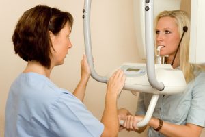
What a CBCT Scan Shows That Regular Dental X-Rays Cannot
A CBCT scan shows three-dimensional details of teeth, jawbone, nerve paths, sinuses, impacted teeth, and some facial structures that regular two-dimensional dental x-rays cannot show as clearly. It is useful when the extra 3D information could change diagnosis, treatment planning, or surgical safety.
CBCT takeaway: A CBCT scan should answer a specific clinical question. Ask what the scan will show, why a regular x-ray is not enough, and how the dental team limits radiation exposure.
What CBCT means
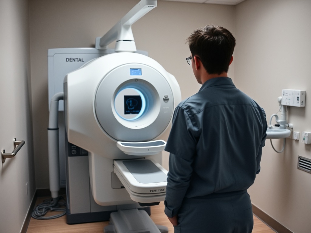
CBCT stands for cone-beam computed tomography. The scanner rotates around the head and uses a cone-shaped x-ray beam to create a three-dimensional image. The FDA’s dental CBCT overview explains that CBCT can reconstruct 3D images of the teeth, mouth, jaw, neck, and related regions.
Regular dental x-rays, such as bitewings or periapical images, are two-dimensional. They are excellent for many routine questions, including cavities between teeth, bone levels, and root areas. But a flat image can overlap structures. CBCT can separate depth, width, and height in ways a regular x-ray cannot.
That does not mean CBCT is better for every visit. It usually gives more information and more radiation than conventional dental x-rays, so the benefit should justify the scan.
What CBCT can reveal
A CBCT scan may help with:
- Implant planning and bone measurement.
- Locating nerves before surgery.
- Evaluating impacted teeth.
- Checking root shape or unusual anatomy.
- Looking at jaw lesions, cysts, or bony changes.
- Assessing some trauma cases.
- Planning certain orthodontic or oral surgery cases.
- Evaluating sinus proximity to upper teeth.
- Understanding failed or complex endodontic cases.
The RadiologyInfo dental cone beam CT resource describes CBCT as useful when regular dental or facial x-rays are not sufficient, including implant placement, impacted teeth, jaw and sinus evaluation, and locating the origin of pain or pathology.
CBCT versus regular dental x-rays
| Feature | Regular dental x-rays | CBCT scan |
|---|---|---|
| Image type | Two-dimensional | Three-dimensional |
| Common uses | Cavities, bone levels, many root and tooth checks | Implants, impacted teeth, complex anatomy, surgical planning |
| Radiation | Usually lower than CBCT | Generally higher than conventional dental x-rays |
| Detail | Excellent for many routine dental questions | Shows depth and spatial relationships |
| Routine use | Common in preventive and diagnostic visits | Used when the 3D answer matters |
| Patient question | “What are you checking?” | “What will this 3D scan change?” |
The ADA’s radiograph guidance emphasizes that dental imaging should be based on clinical need after evaluating the patient, not taken automatically on a fixed schedule. That applies to CBCT too.
Clinical Checks for CBCT Scan Shows That Regular Dental X-Rays Cannot
CBCT can be especially helpful when a dentist needs to know exactly where anatomy sits in three dimensions. For implant treatment, it can show bone shape and nearby structures. For an impacted canine, it can help locate the tooth and its relationship to nearby roots. For some root canal problems, it may show anatomy that a regular image cannot separate.
If you are comparing tooth replacement options, read implant bridges versus individual implants before your consultation. CBCT may be part of that planning, but it should serve the decision, not replace a clear explanation of options.
If orthodontic treatment is being discussed for jaw symptoms or bite strain, the article on orthodontics, TMJ symptoms, and bite strain explains why imaging is only one piece of diagnosis. A scan does not automatically prove that tooth movement will solve pain.
Clinical Checks for CBCT Scan Shows That Regular Dental X-Rays Cannot During Planning
CBCT is powerful, but it has limits. It does not replace a clinical exam, periodontal measurements, symptom history, bite analysis, or medical history. It may not show soft tissues the way a medical CT or MRI can. It also does not decide treatment by itself. Two patients can have similar images and different treatment needs because their symptoms, health, goals, and risks differ.
A CBCT scan can reveal incidental findings, which are unexpected details outside the original dental question. Ask who reviews the scan and what happens if something outside the dental area needs medical follow-up.
What the scan appointment usually feels like
Most CBCT appointments are brief from the patient’s point of view, although the full visit may include positioning, instructions, and image review. You may be asked to remove jewelry, glasses, removable appliances, or anything metal near the head and neck. The team positions your head carefully so the image answers the clinical question. During the scan, you usually stay still while the machine rotates. Tell the team before starting if you have trouble standing, neck discomfort, anxiety in small spaces, or difficulty keeping still.
Radiation questions to ask
You do not need to be afraid of needed imaging, but you should understand why it is being used. Ask:
- What specific question are we trying to answer?
- Could a regular x-ray answer it?
- What field of view will be used?
- How will the settings be adjusted for my size and purpose?
- Will previous images be reviewed first?
- Who interprets the scan?
- Will I receive a copy if I see a specialist?
- How does this scan change the treatment plan?
Pregnant patients should tell the dental team before imaging. A cleaning or urgent dental care may still be appropriate during pregnancy, but imaging decisions should be individualized. The guide on cleanings while pregnant or breastfeeding explains how to discuss pregnancy status before dental visits.
Routine imaging versus urgent diagnosis
A CBCT scan is usually scheduled when a specific treatment or diagnostic question exists. It may be routine within implant or complex oral surgery planning, but it should not feel casual. If you have severe swelling, trauma, fever, or trouble breathing or swallowing, urgent clinical evaluation comes first. Imaging may follow, but the priority is safety and diagnosis.
Use CBCT When the Picture Changes the Plan
CBCT is most useful when three-dimensional anatomy affects treatment choices. Your next step is to ask the dentist what the scan is expected to reveal, why 2D imaging is not enough, and how the result will change the plan you are being asked to consider.
Home Care and Follow-Up for What a CBCT Scan Shows That Regular Dental X-Rays Cannot
Follow-up for What a CBCT Scan Shows That Regular Dental X-Rays Cannot should identify expected changes, symptoms that require a phone call, and the date of the next professional review. Long-term success with What a CBCT Scan Shows That Regular Dental X-Rays Cannot is easier to protect when daily cleaning and maintenance instructions are written clearly as the long-term result is reviewed.